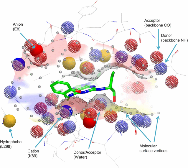
Fig. 1
From: Mapping the 3D structures of small molecule binding sites

SiteHopper patch exemplified for the cofactor binding site of CDK2 (PDB ID: 2A0C) defined by surface protein atoms within 4 Å of a bound ligand (shown in green). A pharmacophore model defines pseudocenters for five key interaction types: hydrogen bond donor (blue mesh), hydrogen bond acceptor (red mesh), anion (red solid), cation (blue solid) and hydrophobe (yellow). Surface vertices also encode the shape of the binding site (gray). Image produced using VIDA [17]